Cranial Nerve & Foramina 3D Model
Project Overview
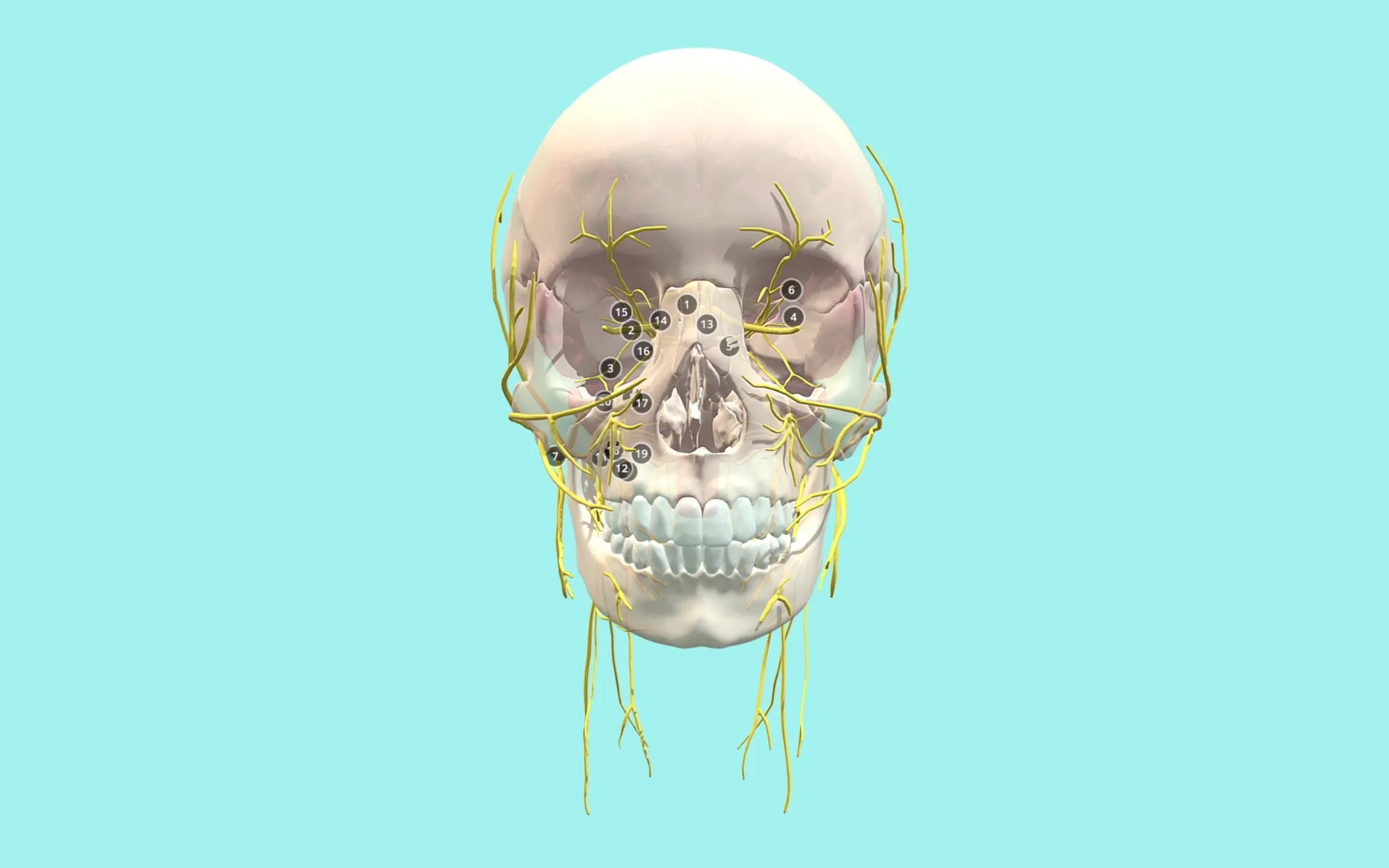
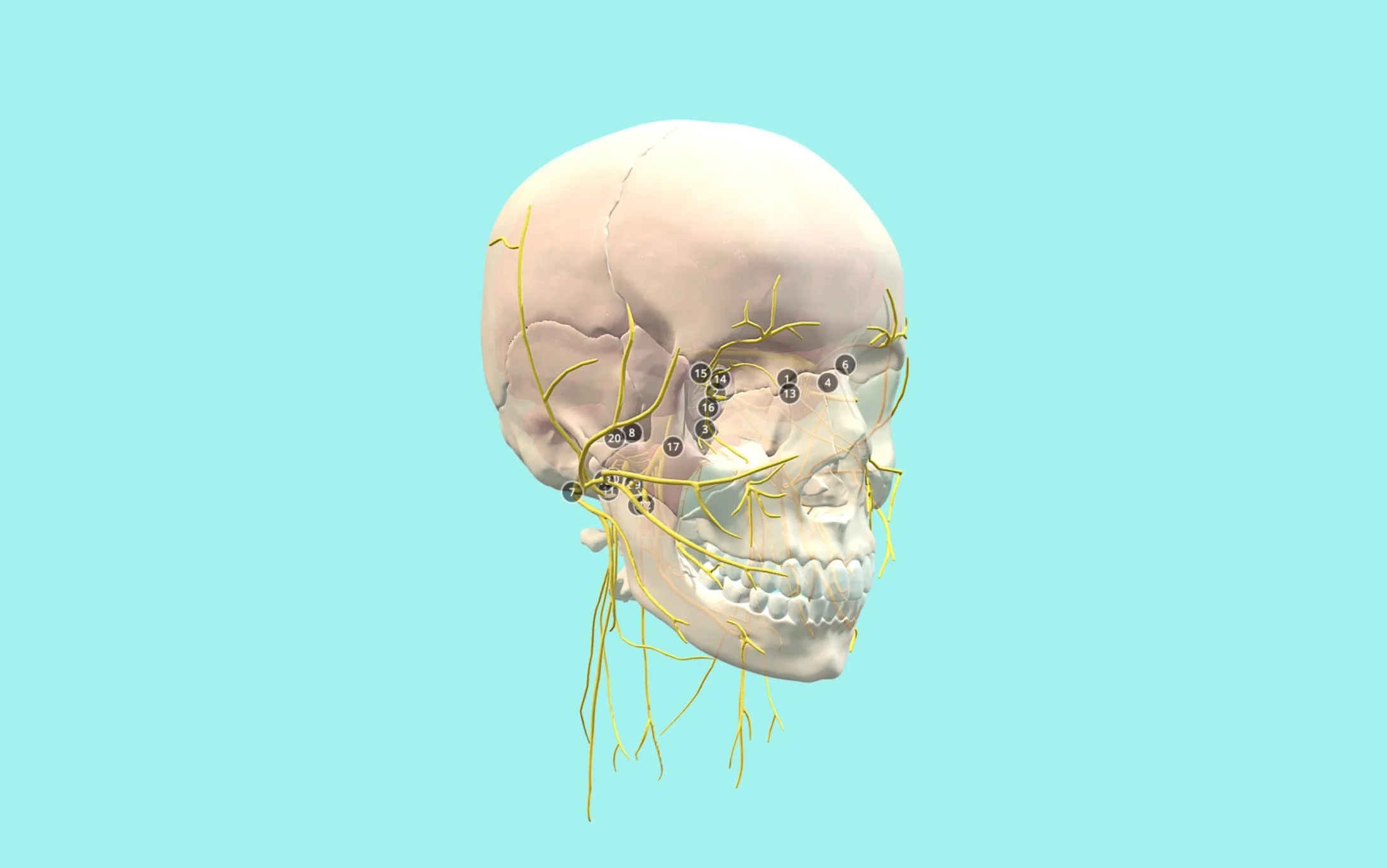
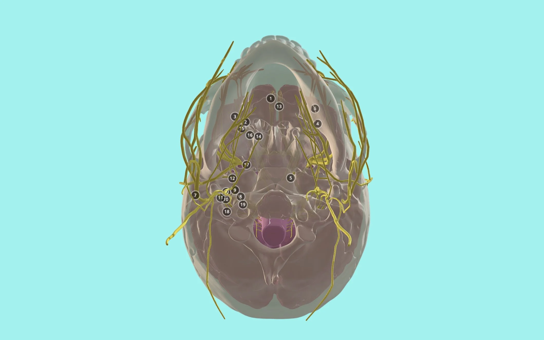
The cranial nerves follow complex pathways through openings in the base of the skull known as foramina. While completing cadaver dissections, I realized how difficult it could be to see where each cranial nerve originated in the brain and where it exited the skull, as the surrounding tissues and structures often obscured these relationships. I created this interactive 3D model for the University of Dundee to isolate the key anatomy and help students clearly understand the origins, pathways, and exit points of the twelve cranial nerves.
Creating the Model
I began with a skull base from the BodyParts3D dataset and refined it. I created a brain and added the cranial nerves to create a clear and accurate educational model. Each nerve pathway was carefully reviewed and positioned so that its relationship with the skull and associated foramina could be understood from multiple angles.
The nerves were given a contrasting color and the skull was made partially transparent, allowing structures that would normally be hidden inside the cranium to remain visible. Numbered annotations were then added to identify important openings and anatomical relationships.
The Final Interactive Model
The completed model was prepared for Sketchfab, where students can rotate, zoom, and explore the anatomy from any direction. This interactive format makes it easier to understand the three-dimensional relationships between the cranial nerves and the openings through which they pass.
Explore the Final Model
Explore the final model below by rotating the skull, zooming in, and viewing the cranial nerves from different angles. Use the interactive labels to follow each nerve from its origin in the brain to the opening where it exits the skull.