Embryology Animation & E-Learning Resource
This educational resource was created to help university students understand the complex folding and development of the human embryo. The project combines anatomical research, 3D modeling, medical animation, voice-over, and website design to present embryology in a more visual and engaging format.
I developed the entire project from its initial research and concept through to the finished 3D models, animations and e-learning website. The resource was designed to supplement embryology teaching at the University of Dundee and the University of St Andrews.
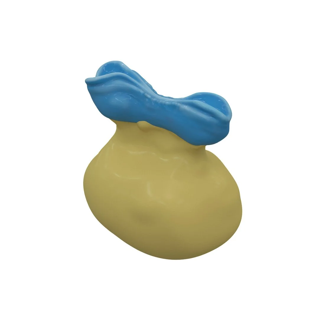
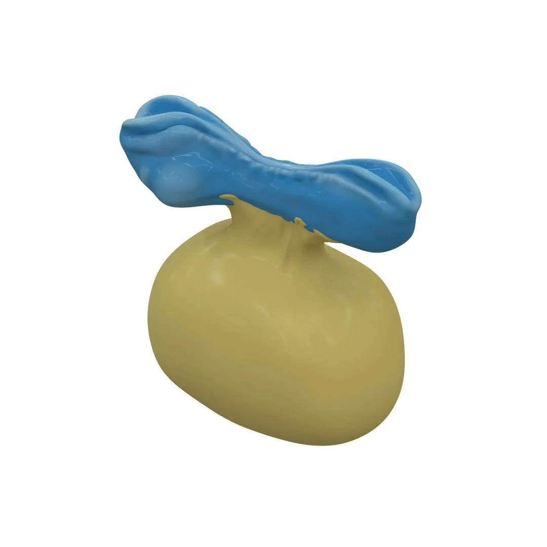
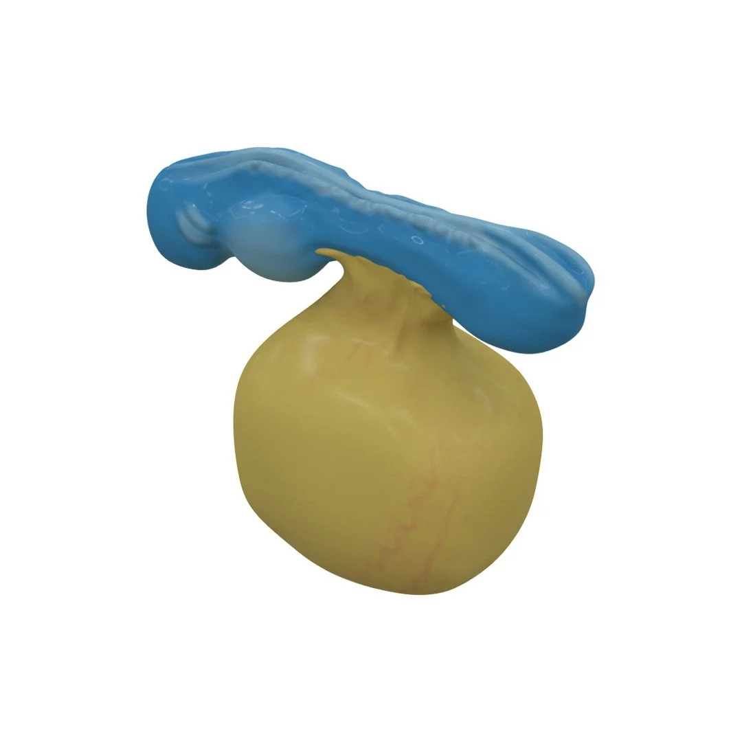
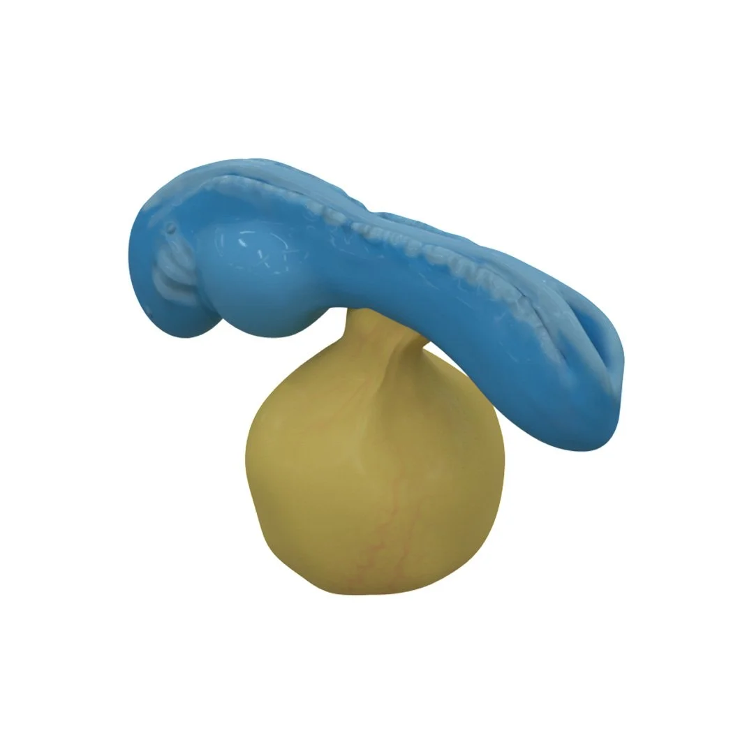
Week 4 3D Models
-
![Day 22]()
Day 22
-
![Day 23]()
Day 23
-
![Day 24]()
Day 24
-
![Day 25]()
Day 25
-
![Day 26]()
Day 26
-
![Day 27]()
Day 27
-
![Day 28]()
Day 28
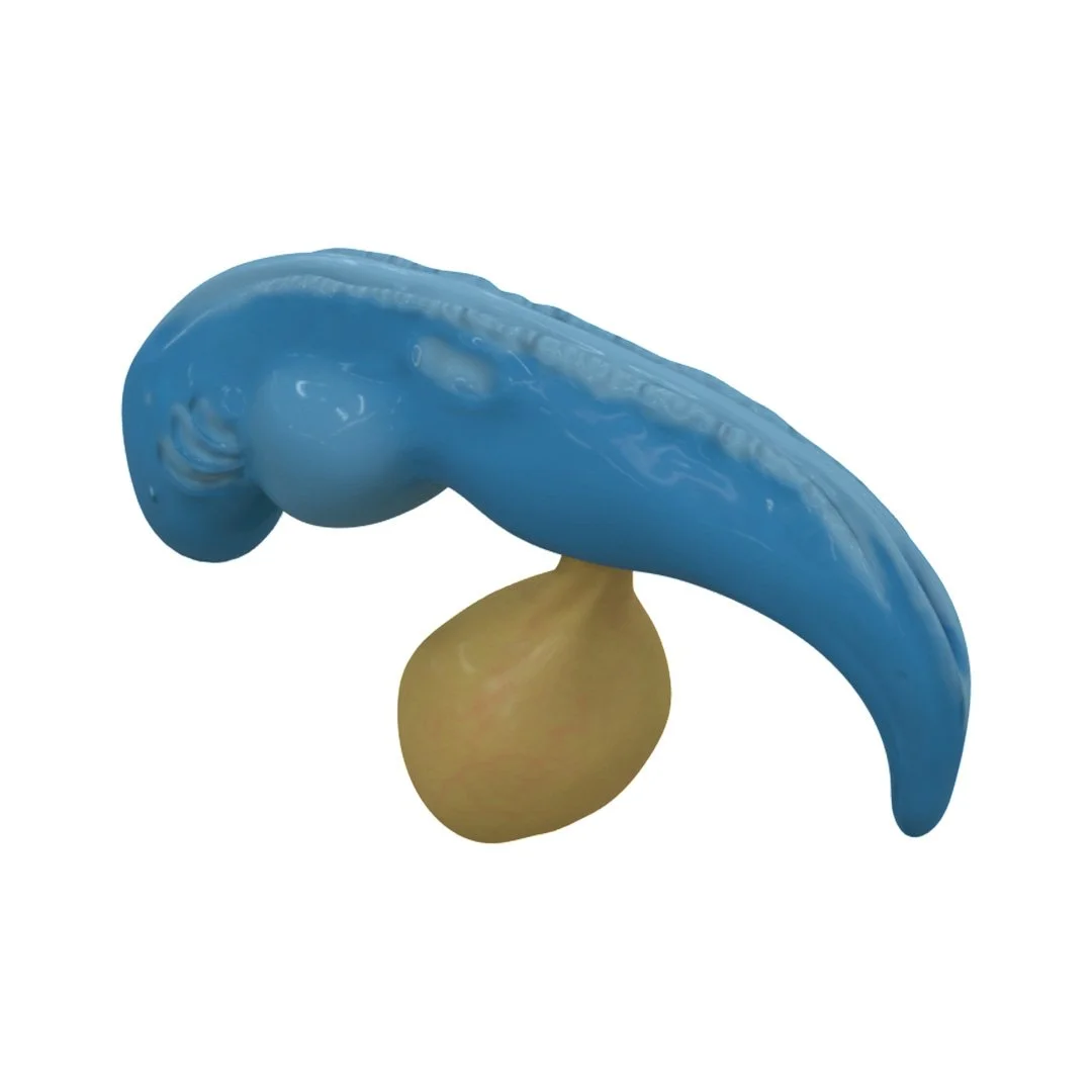
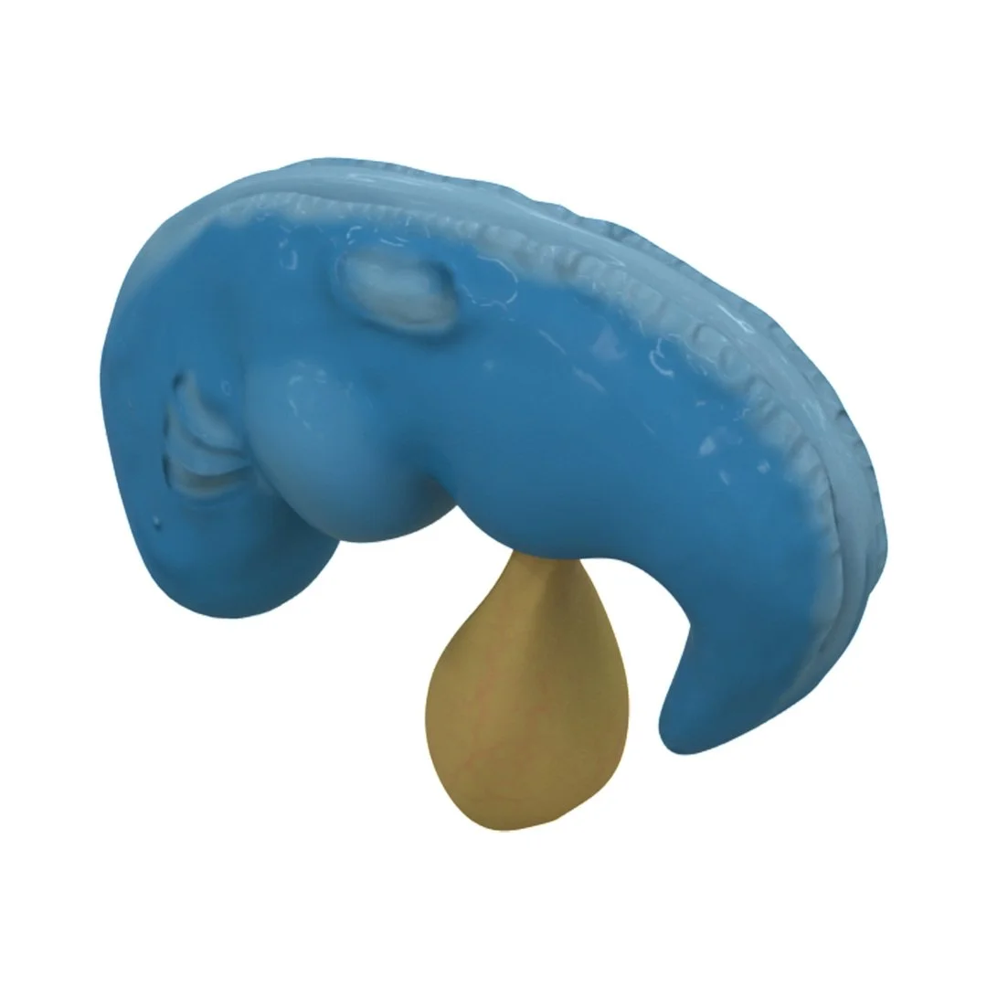
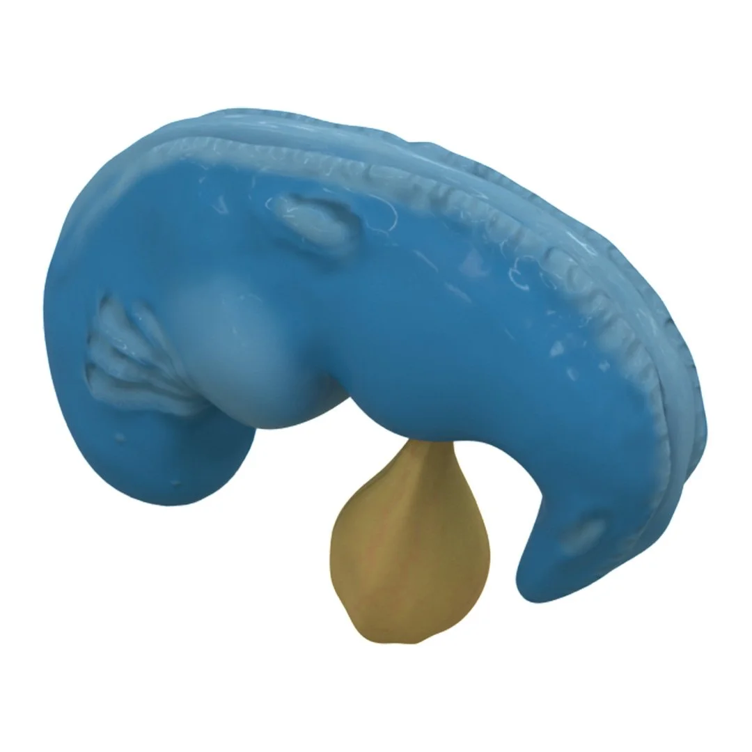
The Challenge
Embryology is traditionally taught using textbook illustrations, lecture slides, and written descriptions. Although these resources can show individual stages of development, it can be difficult for students to understand how the embryo transitions from one stage to the next.
During the fourth week of development, the embryo folds in several directions at the same time. This transforms the relatively flat embryonic disc into a three-dimensional form. Understanding this process requires students to interpret multiple two-dimensional images and mentally reconstruct a complex sequence of movement.
The goal of this project was to make that transition easier to visualize by showing the embryo developing continuously through animation and from multiple viewpoints.
Anatomical Research
The project began with extensive research into the shape and anatomy of the developing human embryo.
Because no single reference clearly documented every structure and stage from all of the angles required for the animation, I compared information from embryology textbooks, research papers, diagrams, photographs, and existing physical and digital models. I collated these sources to determine how the embryo’s external form and internal structures should appear at each stage of development.
This research was particularly important when interpreting how structures changed between days. The final models needed to be scientifically accurate while also remaining clear enough for students who were encountering the subject for the first time.
3D Modeling and Medical Animation
I sculpted the embryo models in ZBrush, creating each developmental stage and refining the anatomical structures as the embryo changed in shape. I then added color and surface texture to distinguish important structures and make the models easier to interpret.
The models were transferred into Autodesk Maya, where I created the animation showing the embryo folding and developing between days 22 and 28. Rather than presenting the stages as isolated models, the animation demonstrates the movement and transformation that occurs between them.
I created both cranial-caudal and lateral views so students could observe the folding process from different perspectives and develop a stronger three-dimensional understanding of the anatomy.
The animations were then assembled in Adobe After Effects, where I added labels, explanatory text, timing, transitions, and voice-over narration.
The Final 3D Animation
The final animation follows the folding and development of the human embryo from days 22 to 28. By showing the process continuously from cranial-caudal and lateral views, it helps students understand how the embryo changes from a relatively flat structure into a complex three-dimensional form. Labels, narration, and color-coded anatomy guide the viewer through each stage of development. The animation was supported by a simple e-learning website that provided additional context, terminology, and practice questions.
Final 3D animation
Designing the E-Learning Experience
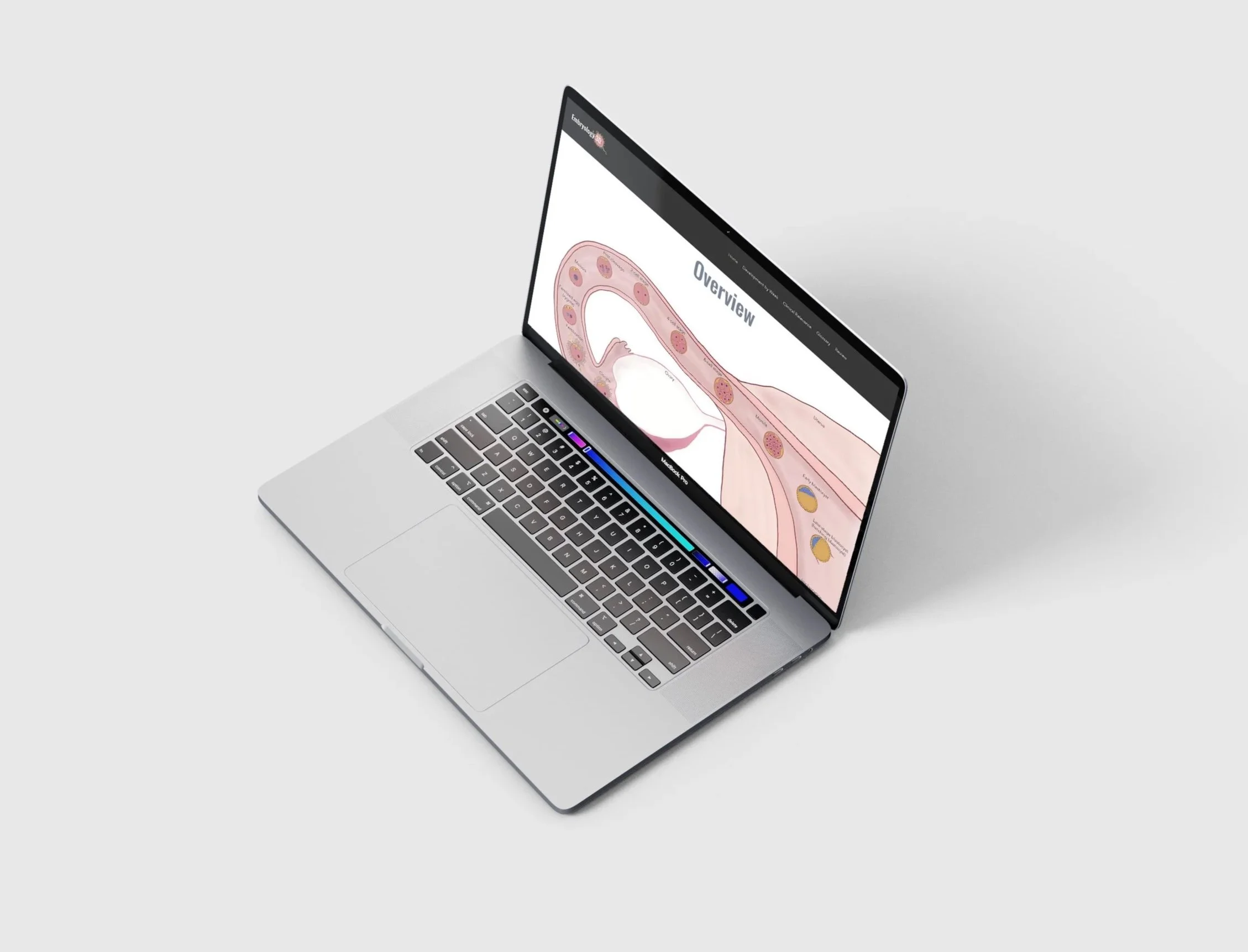
The animation became the central feature of a dedicated embryology learning website. I designed the site to give students control over how they moved through the material, allowing them to pause, replay, and revisit the animation as often as necessary.
The website also included:
Supplemental explanations of each developmental stage
Individual 3D models showing key stages of development
A glossary of common embryology terminology
Clinical information connecting development with relevant conditions
Review questions to help students test their understanding
Bringing these elements together created a more complete learning experience than an animation alone. Students could first observe the developmental process, then explore individual stages and reinforce their understanding through supporting text and questions.
EMBRYOLOGY 3D
The Final Resource
The finished project transformed a complex, highly spatial process into a structured visual learning experience. By combining scientific research, anatomically accurate 3D models, animation, narration, and interactive educational content, the resource was designed to help students understand not only what the embryo looks like at each stage, but how and why its shape changes over time.
My role: Research, medical illustration, 3D modeling, texturing, animation, compositing, narration, educational content, and website design
Software: ZBrush, Autodesk Maya, Adobe After Effects, Adobe Photoshop, Adobe InDesign and Wix