From Carbon Dust to Pixels: The Evolution of Medical Art
For centuries, understanding the human body was a dangerous, often illegal pursuit involving midnight dissections and smuggled sketches. We’ve come a long way from the 'muscle men' of the Renaissance. Today, medical illustration is no longer about just recording death, it’s about visualizing life. This post is a look at the pioneers who turned a macabre craft into the field of medical illustration we know today. If you want more background history, read this post first and then come back!
Another quick note, before getting started. This post contains affiliate links. This means I may earn a commission should you chose to make a purchase using my link.
The Fathers of Modern Illustration: Brödel and Netter
Brödel:
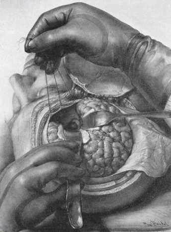
As medicine became more clinical and technology advanced, medical art became more specialized. In the early 20th century, Max Brödel (1870-1941), born in Leipzig, Germany, founded the first department of medical illustration at Johns Hopkins University in the US and is known as the "father of modern medical illustration”. He created new techniques using carbon dust that gave tissues depth and texture. He worked closely with surgeons, especially Dr. Howard Kelly, to create first-of-their-kind illustrations showing the surgeon’s point of view. Illustrations like this became paramount for learning surgical techniques and enhanced surgeons’ understanding. His carbon dust technique is still taught in medical art courses today!
Netter
Often called the "Medical Michelangelo" for his gorgeous work, American surgeon and illustrator Frank Netter (1906–1991) created a legacy that remains the gold standard for medical education. Early in his career, Netter began illustrating to supplement his income, but his work was so well-received that it soon became his full-time calling. His 1948 debut, The CIBA Collection of Medical Illustrations, featured over 4,000 pieces and paved the way for the iconic Netter’s Atlas of Human Anatomy.
To this day, the Atlas is a rite of passage for medical students. Every medical illustrator should have a copy of his atlas for reference or just to use as inspo! He’s one of my favorite medical illustrators of all time because his work is accurate, beautiful and seems to be as simplified as possible without losing pertinent details. Basically his work is perfect? If you’re as obsessed as I am, check out this biography by netterimages.com to see his full life story!
The beauty of human anatomy, captured by the master himself. Take a peek inside Netter’s Atlas, the ultimate reference for turning an obsession with the human body into a professional career.
An illustration by me (above): I channeled my inner 'Medical Michelangelo' for this one. Frank Netter’s influence is everywhere in medical art, and for this illustration, I focused on his signature style of using vibrant colors and clean lines to give the work that extra bit of polish that really makes it stand out. It’s a rewarding challenge to take those mid-century master techniques and apply them to a modern digital canvas.
The Digital Frontier: From Paper to Pixels
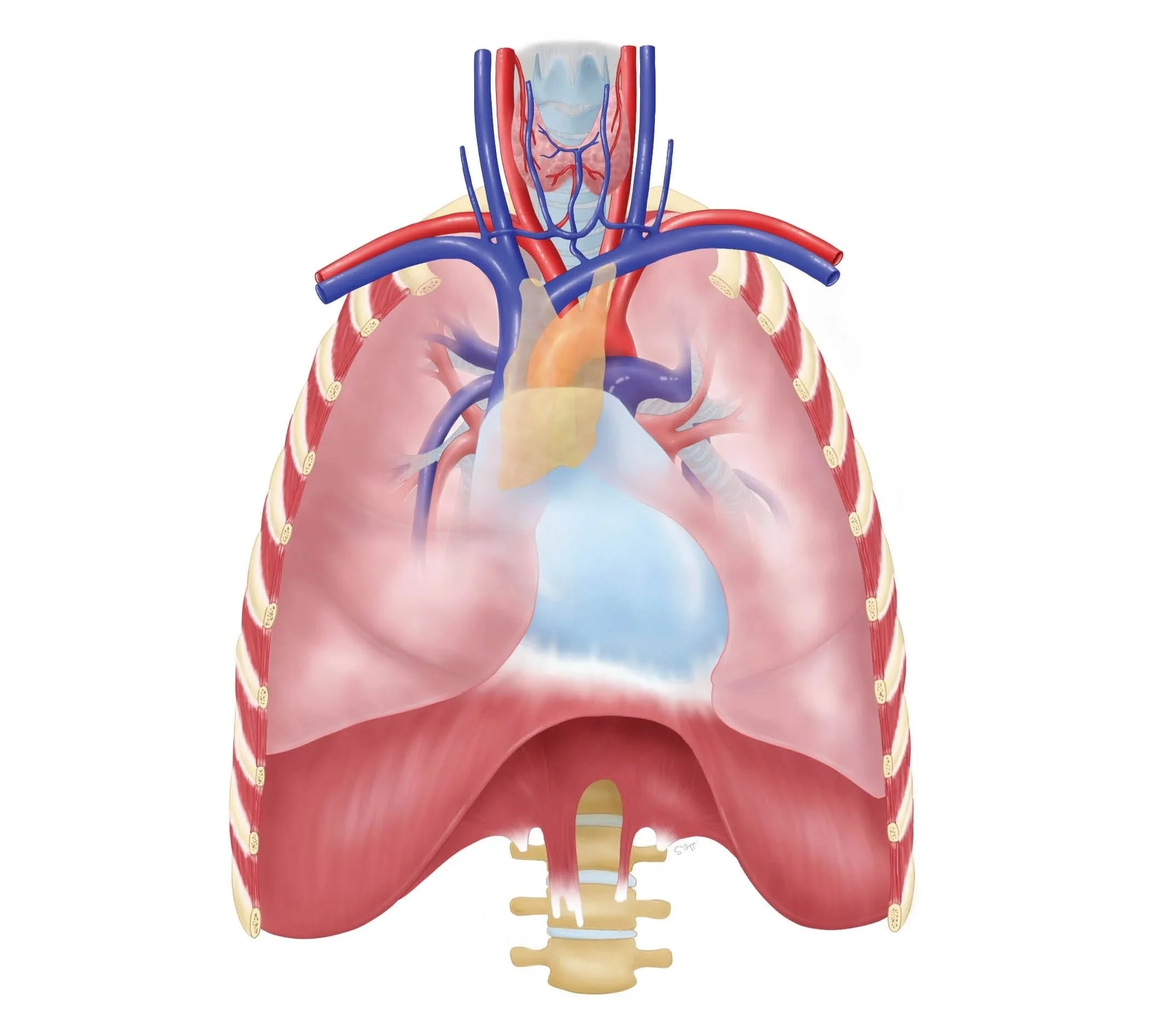
Today, we are living in a second Renaissance. We’ve come from Galen’s four humors to mapping individual cells under high-powered microscopes. The field of medical art has evolved from ink and parchment to digital illustration, animation, 3D modeling, virtual reality (VR), and augmented reality (AR). It’s INCREDIBLE! There are so many ways to "do” medical art now!
Modern illustrators aren't just drawing; they are building interactive worlds. We can now "walk through" a human heart in VR or watch a 3D animation of a molecular drug binding to a receptor. As technology evolves, so does our ability to visualize the invisible.
Beyond the "cool factor," these advancements are the backbone of health literacy. In the past, medical knowledge was locked away in Latin texts for the elite; today, medical art acts as a universal language. I love that my work helps patients to understand their own body. When a patient can see an animation of their own surgery or a 3D model of how their digestive system works, it bridges the gap between complex science and personal well-being. By making the invisible visible, we empower people to make informed decisions about their own health.
The journey from carbon dust to virtual reality shows just how far we’ve come in our quest to understand ourselves. I’m excited to see where the next 'Digital Renaissance' takes us. Thanks for geeking out on medical art history with me!
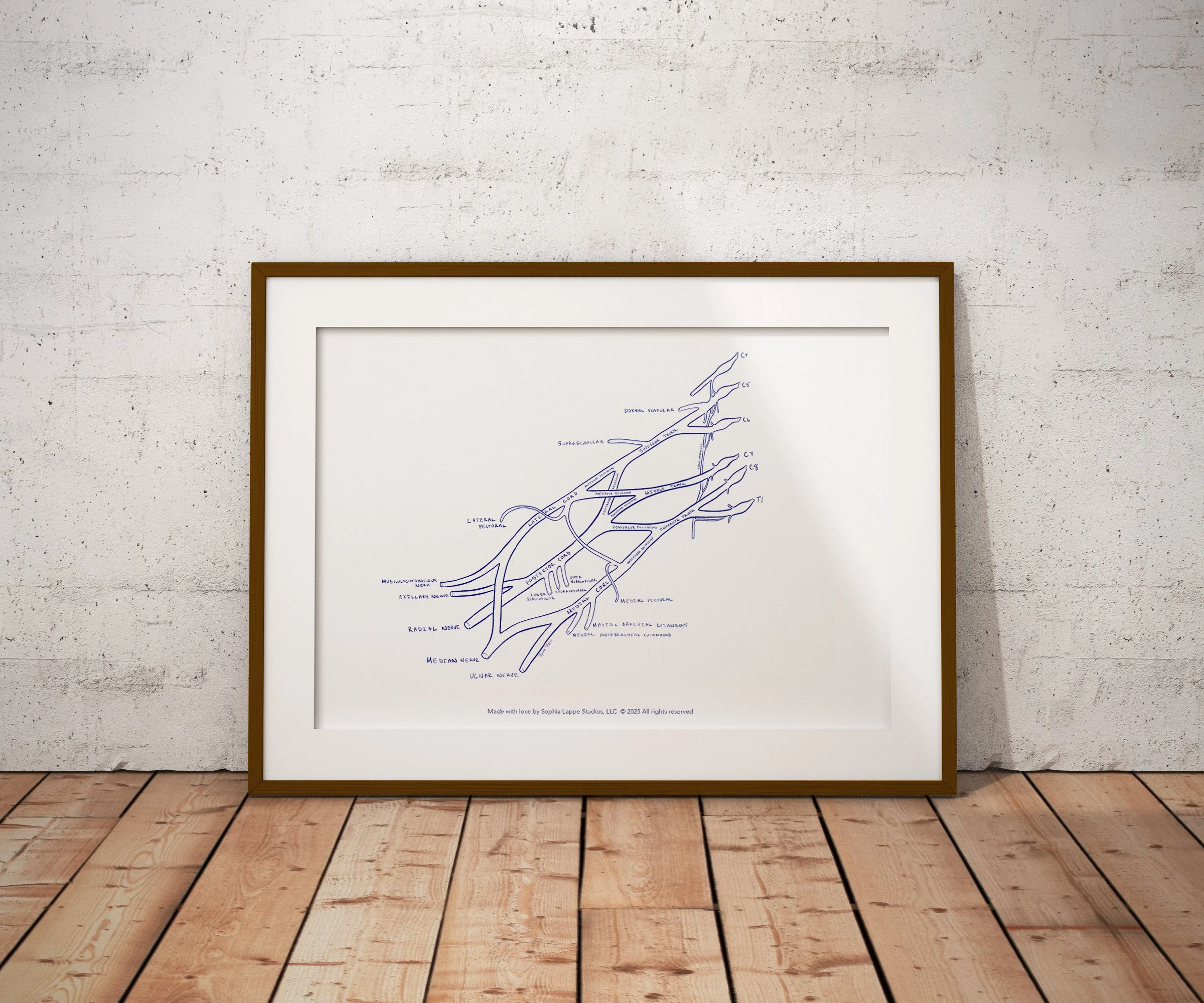
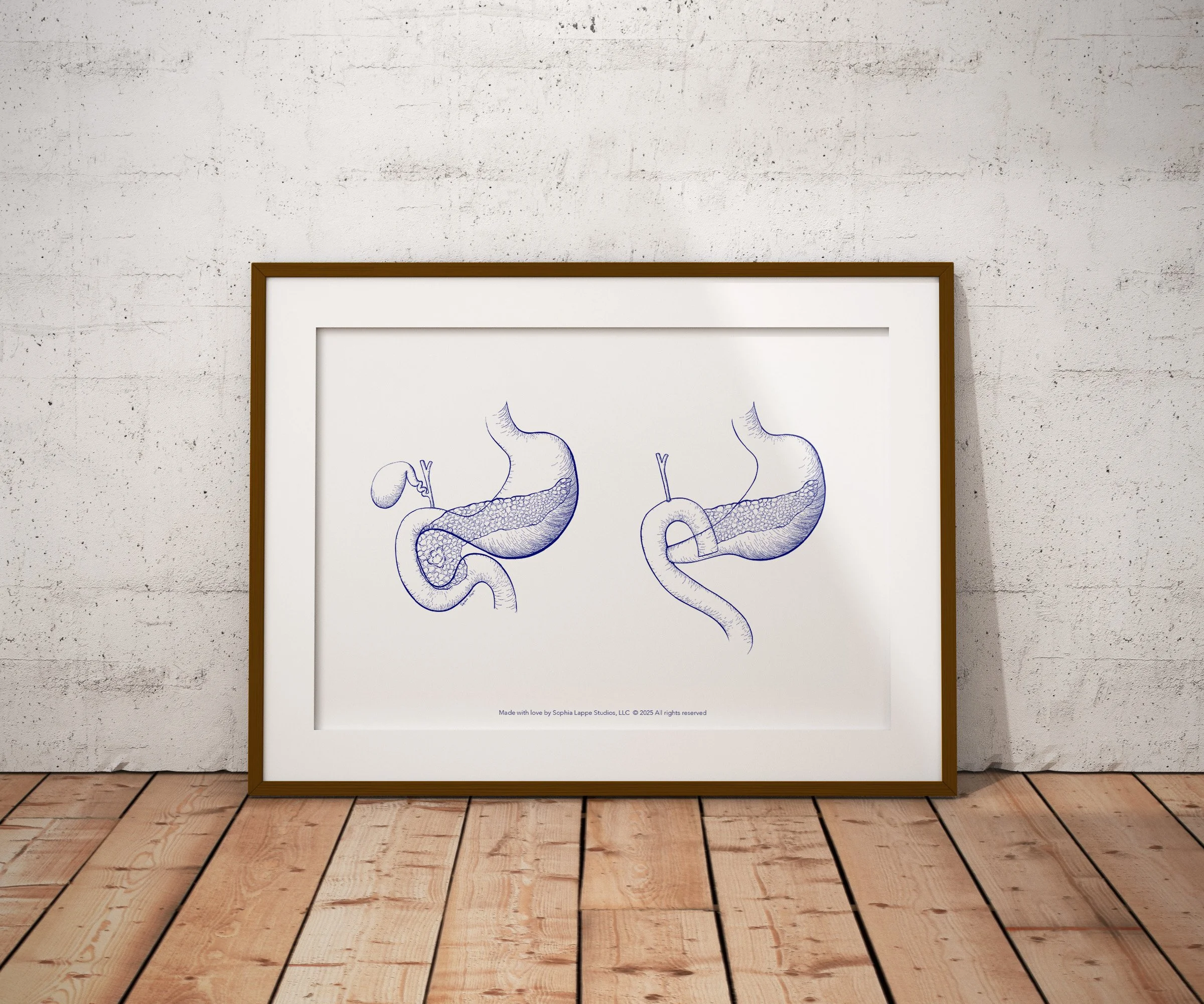
If you share my love of anatomy, I’ve turned some of my favorite illustrations into high-quality prints. You can find them right here in my shop.

If you’d like to work together, have questions or need advice about medical art, please don’t hesitate to get in touch!