Drawing the Dead: How the Open Body Moved from Mystery to Masterpiece
From Taboo to Textbook: The Dawn of Anatomy
It used to be unthinkable to cut someone open after they died just to explore their insides. And then, to request to draw their cold, lifeless body? Forget about it! So, how did this previously taboo topic come to lend itself to a whole career?
It took a lot of bold and curious people to get to the point we’re at today where students can learn anatomy hands on (thanks to all of the people who have graciously donated their bodies to science). But learning anatomy through dissection wasn’t always this acceptable or accessible. There is a long and very dark history of grave robbers, serial killers, and murderers working to source bodies for doctors… but that’s for another post! This blog post is a brief overview of the advancements in anatomy and medical illustration throughout history.
Ancient Origins: Papyri, Silk, and Early Observation
Let’s start about 4,600 years ago in Ancient Egypt with a man named Imhotep. He was a statesman, architect, and healer associated with the Pharaoh Djoser in the 27th century BCE and was later worshiped as a god of medicine. Imhotep is sometimes linked to the Edwin Smith Papyrus. Copied around 1600 BCE from an older source, the papyrus is best known as the oldest surviving surgical treatise and one of the earliest Egyptian medical texts. It is especially notable for its structured approach to observation, diagnosis, and treatment, as well as its detailed descriptions of internal anatomy and remedies for a range of injuries and ailments. This document fundamentally changed how medicine was viewed because it relied on observation over magic. It is primarily a medical text, however, not a surviving illustrated medical atlas.
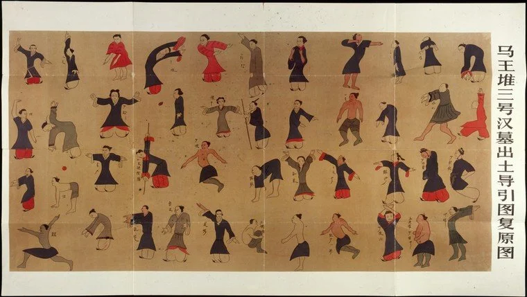
Further east, the manuscripts found in the Mawangdui Tomb 3, sealed in 168 BCE, include important early Chinese medical materials and the illustrated Daoyin tu that show how people in Ancient China understood the body. Some of these works show anatomical charts and diagrams of the acupuncture meridians. These works are foundational for the history of Chinese medicine, but they are not always classified as the oldest “medical illustrations”, because some are textual medical manuscripts and others are better understood as exercise or medical charts. But I think it’s important to include both of these texts, the Mawangdui silk manuscripts and the Edwin Smith Papyrus, to show how different cultures around the world understood, practiced and documented medicine.
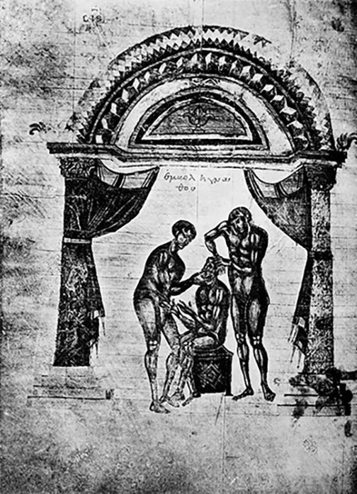
Many histories of medical illustration trace the earliest and first true instructional medical illustration to Hellenistic Alexandria in the 4th or early 3rd century BCE. The figures were often portrayed in a slightly squatting position, a pose designed to display the muscles and joints clearly. From surgeries to the details of obstetrics and medicinal plants, these papyri represent when medical art became a teaching tool. For the first time, a healer didn't just have to hear about anatomy; they could finally see it.
The Foundation of Western Medicine: Humors and Early Dissection
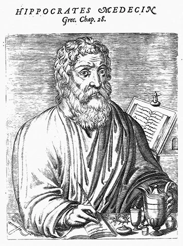
Hippocrates of Kos
Hippocrates of Kos, a Greek physician, helped shift Greek medicine toward natural explanations of disease and observation based medicine. In the Hippocratic tradition, illness was understood as arising from natural causes like environmental factors, lifestyle and diet, rather than from superstition or angry gods. Hippocratic medicine was traditionally associated with the four humors of the body (black bile, yellow bile, blood, phlegm) which held that disease arose when there were imbalances within the body. Although Hippocrates himself is not closely tied to anatomical illustration or human dissection, his emphasis on observation and natural causes laid important groundwork for later Greek physicians, including the Alexandrian anatomists who advanced dissection-based study of the body.
Herophilus and Erasistratus
Hellenistic Alexandria was a crucial center for anatomists because most human dissection was considered too taboo or banned in other places at this time. It was here that Herophilus of Chalcedon (335–280 BC), considered the father of anatomy, carried out systematic dissections of human cadavers and made major discoveries about the brain, nerves and other organs. Notably, he argued that the brain was the center of intelligence and helped advance the study of the nervous system by distinguishing sensory from motor nerves. His contemporary, Erasistratus of Chios (304-250 BC), considered the father of physiology, focused on the function of the organs, especially the heart. Herophilus and Erasistratus established the first medical school in Alexandria and both made lasting contributions to the field of medicine through dissection and careful observation.
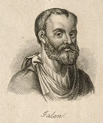
Galen
Galen was a Greek physician who worked in the Roman Empire and served several prominent Roman rulers, including Marcus Aurelius and Commodus. His understanding of the body was influenced by the Hippocratic tradition of the four humors. Galen further developed this theory by linking the humors to human temperaments: blood (sanguine), black bile (melancholic), yellow bile (choleric,) and phlegm (phlegmatic). He also made major contributions to the understanding of the circulatory system especially with his observations in the differences between venous and arterial blood, helping show that arteries carry blood rather than air. Because human dissection became largely restricted or taboo in Roman society, Galen based much of his anatomical work on dissections and vivisections (live dissection) of animals, especially pigs and monkeys. It remains unclear whether Galen himself created illustrations to accompany his manuscripts or whether such images were added later as his texts were copied and transmitted.
The Renaissance Revolution: Vesalius Reclaims the Body
As we move into the Renaissance, we hit a massive turning point with Andreas Vesalius. Before him, many still clung to Galen’s animal-based observations, but Vesalius changed the game by performing his own human dissections. His 1543 masterpiece, De Humani Corporis Fabrica Libri Septem (On the Fabric of the Human Body), showed human anatomy in ways the world had never seen.
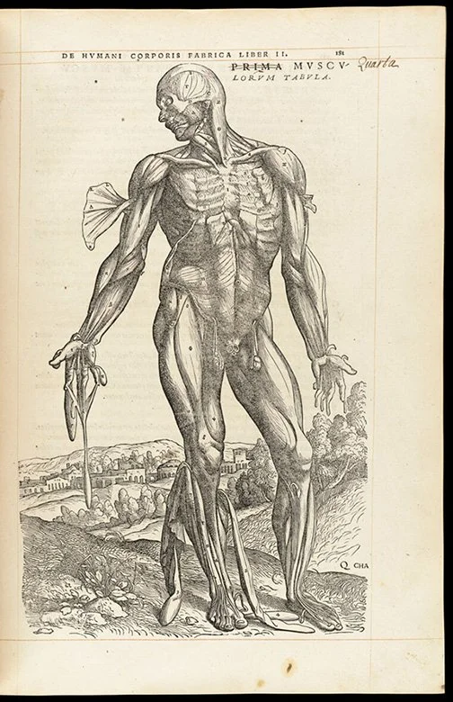
What makes Vesalius’s work so iconic isn’t just the anatomical accuracy, but the artistry. And this is where I could really start to geek out. He depicted "muscle men" in contrapposto poses, standing naturally against lush Italian landscapes, making the macabre study of death feel vibrant and alive. This is so important for the history of medical art because after over 1000 years of banned or taboo human dissections people were not used to seeing the inside of a body. So, Vesalius, showing great understanding of his audience and human anatomy, went to extensive lengths to make his work more palatable to the people. He is intelligent and thoughtful, you’ve gotta love him!
The Macabre Business of the 1800s
By the 19th century, the demand for medical knowledge outpaced the legal supply of bodies. This led to a grisly era of resurrectionists, grave robbers who dug up the recently deceased to sell to medical schools. In the most extreme cases, like the infamous Burke and Hare in Scotland, serial killers actually murdered people to keep up with the doctors' demands. Scary times!!!
Because fresh cadavers were so scarce and decomposed quickly, dissections became a public spectacle. They were performed in anatomical theaters like this one, where students and even some curious members of the public sat in steep, circular rows to watch a professor work. In this high-pressure environment, the need for permanent, accurate medical illustrations became more desperate than ever.
Stay tuned for part 2 where we move from the graveyard to the digital frontier!
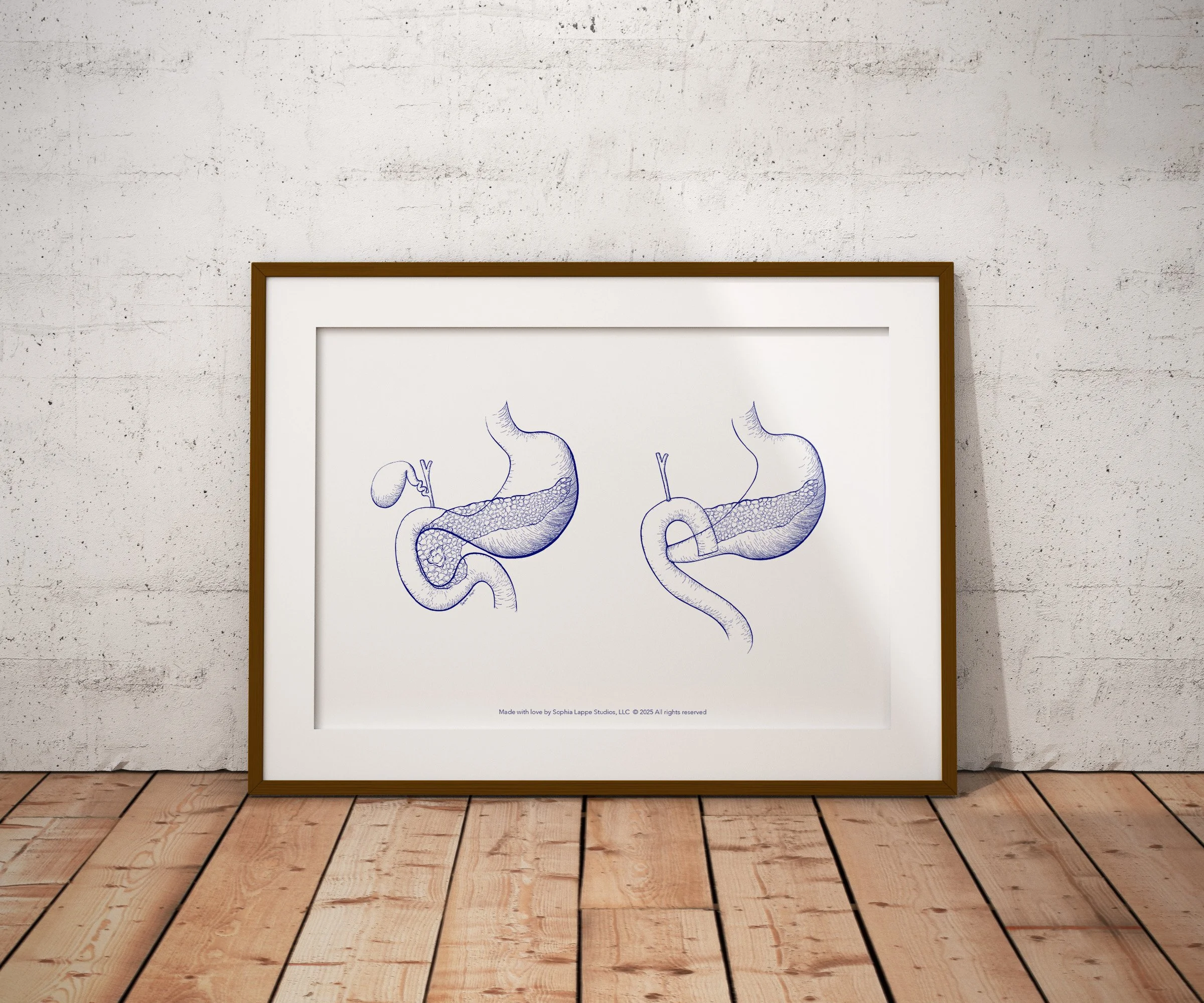
If you share my love of anatomy, I’ve turned some of my favorite illustrations into high-quality prints. You can find them right here in my shop.

If you’d like to work together, have questions or need advice about medical art, please don’t hesitate to get in touch!